Understanding Mycosis Fungoides: Symptoms, Diagnosis, and Treatment Explained
Mycosis fungoides is a rare type of T-cell lymphoma that primarily affects the skin. As the most common form of cutaneous T-cell lymphoma, this condition typically develops slowly and can be challenging to diagnose in its early stages. Understanding its symptoms, diagnosis methods, and treatment options is crucial for both patients and healthcare providers.

What is Mycosis Fungoides and How Does it Develop?
Mycosis fungoides originates from a malignant transformation of T-helper cells, a specific type of white blood cell that normally helps coordinate immune responses. These cancerous cells initially accumulate in the skin, creating the characteristic patches and lesions associated with the condition. The exact cause of this cellular transformation remains unclear, though researchers believe it involves a combination of genetic predisposition and environmental factors.
The disease typically develops through three distinct stages. The patch stage represents the earliest phase, where thin, scaly, and often itchy red patches appear on the skin. These patches frequently occur in areas protected from sun exposure, such as the buttocks, hips, and lower trunk. The plaque stage follows, characterized by thicker, raised lesions that may ulcerate. In advanced cases, the tumor stage develops, featuring large nodules and potential spread to lymph nodes and internal organs.
What Are the Key Symptoms of Mycosis Fungoides?
The symptoms of mycosis fungoides vary significantly depending on the stage and extent of the disease. Early-stage symptoms often mimic common skin conditions like eczema or psoriasis, contributing to diagnostic delays. Patients typically experience persistent, itchy red patches that may appear scaly or flaky. These patches often have irregular borders and can vary in size from small spots to large areas covering significant portions of the body.
As the condition progresses, symptoms become more pronounced and distinctive. The skin lesions may thicken and develop into raised plaques that feel firm to the touch. Some patients experience hair loss in affected areas, particularly on the scalp. Advanced stages may present with tumor-like nodules that can ulcerate and become infected. Systemic symptoms such as unexplained weight loss, fever, and enlarged lymph nodes may indicate disease progression beyond the skin.
How is Mycosis Fungoides Diagnosed?
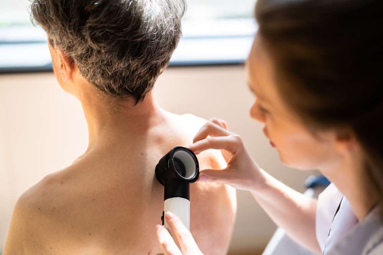
Diagnosing mycosis fungoides requires a comprehensive approach combining clinical examination, laboratory tests, and specialized procedures. The diagnostic process often begins with a thorough dermatological evaluation, where healthcare providers assess the appearance, distribution, and characteristics of skin lesions. However, visual examination alone cannot definitively diagnose the condition due to its similarity to other skin disorders.
Skin biopsy remains the gold standard for diagnosis, involving the removal of a small tissue sample for microscopic examination. Pathologists look for characteristic changes in skin architecture and the presence of atypical T-cells. Multiple biopsies may be necessary, as early-stage lesions can be difficult to distinguish from benign inflammatory conditions. Additional tests include immunohistochemistry to identify specific cell markers and molecular studies to detect T-cell receptor gene rearrangements.
Advanced diagnostic techniques may include imaging studies such as CT scans or PET scans to evaluate potential spread to lymph nodes or internal organs. Blood tests can assess overall health status and detect circulating malignant cells in advanced cases. Flow cytometry of peripheral blood may identify abnormal T-cell populations, providing additional diagnostic information.
Treatment Approaches and Management Strategies
Treatment for mycosis fungoides depends heavily on the stage of disease and the extent of skin involvement. Early-stage disease typically responds well to skin-directed therapies, while advanced stages may require systemic treatments. Topical therapies include corticosteroids, nitrogen mustard, and retinoids, which can effectively control symptoms and reduce lesion size in many patients.
Phototherapy represents another important treatment modality, particularly ultraviolet B (UVB) and psoralen plus ultraviolet A (PUVA) therapy. These treatments can achieve significant improvement in skin lesions while maintaining good quality of life. Radiation therapy may be used for localized areas of disease or as palliative treatment for advanced lesions.
Systemic therapies become necessary for advanced-stage disease or when skin-directed treatments prove insufficient. Options include interferon-alpha, retinoids, chemotherapy agents, and newer targeted therapies. Stem cell transplantation may be considered for younger patients with advanced disease who have suitable donors.
Prognosis and Long-term Outlook
The prognosis for mycosis fungoides varies considerably based on the stage at diagnosis and response to treatment. Early-stage disease generally carries an excellent prognosis, with many patients experiencing normal or near-normal life expectancy. The 10-year survival rate for stage I disease exceeds 80%, while more advanced stages have correspondingly lower survival rates.
Factors influencing prognosis include age at diagnosis, extent of skin involvement, presence of blood involvement, and response to initial treatment. Regular follow-up care is essential for all patients, as the disease can progress over time and may require treatment modifications. Patient education about skin self-examination and recognition of concerning changes plays a crucial role in long-term management.
Understanding mycosis fungoides empowers patients and families to recognize potential warning signs and seek appropriate medical evaluation. While this rare form of lymphoma presents significant challenges, advances in diagnostic techniques and treatment options continue to improve outcomes for affected individuals. Early recognition and proper medical management remain key factors in achieving the best possible results for patients diagnosed with this complex condition.
This article is for informational purposes only and should not be considered medical advice. Please consult a qualified healthcare professional for personalized guidance and treatment.


